In our formulation development process, we rely on human in vitro models of various tissues as an alternative to animal models to test our drug delivery systems. While these models may have limitations in terms of complexity, we believe that using human cells offers several advantages over animal testing when it comes to tailoring drug delivery systems to human biology. Therefore, our research is primarily focused on in vitro models based on human cells and tissues, striking a balance between the required biological intricacy and the ability to conduct valid and predictive testing of new medicines. To meet specific scientific requirements, our models span a range of complexities, from single-cell studies and cell monolayers to intricate three-dimensional organotypic cultures. These models incorporate multiple types of human cells, as well as ex vivo tissue biopsies. We believe that this approach allows us to capture key aspects of tissue behavior and response to drug delivery systems.
Modular In vitro Models
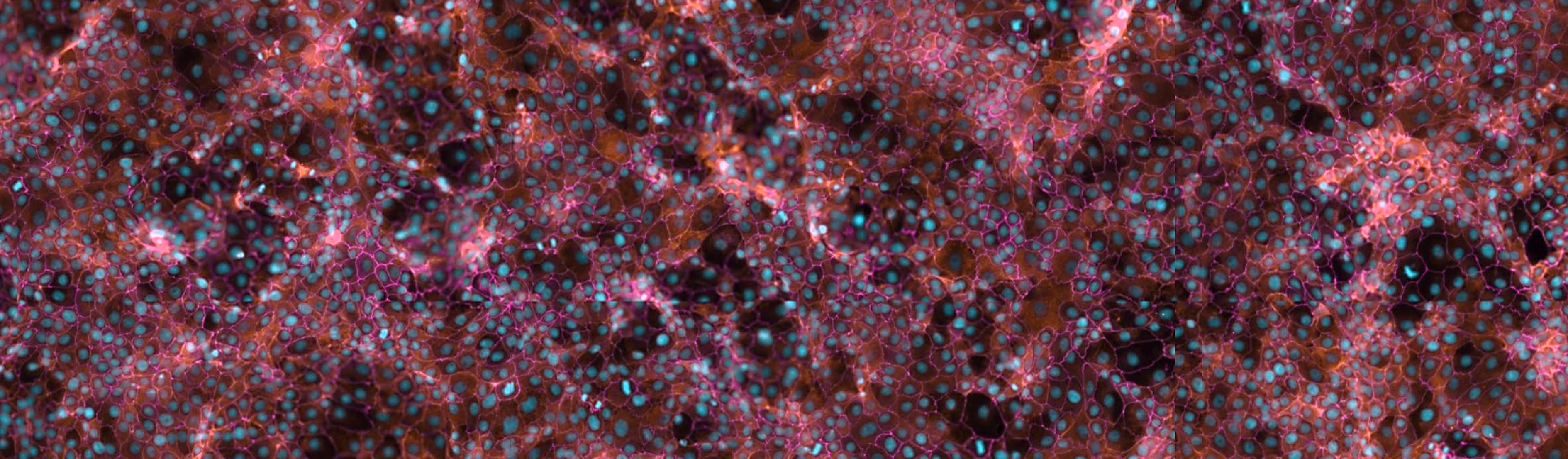
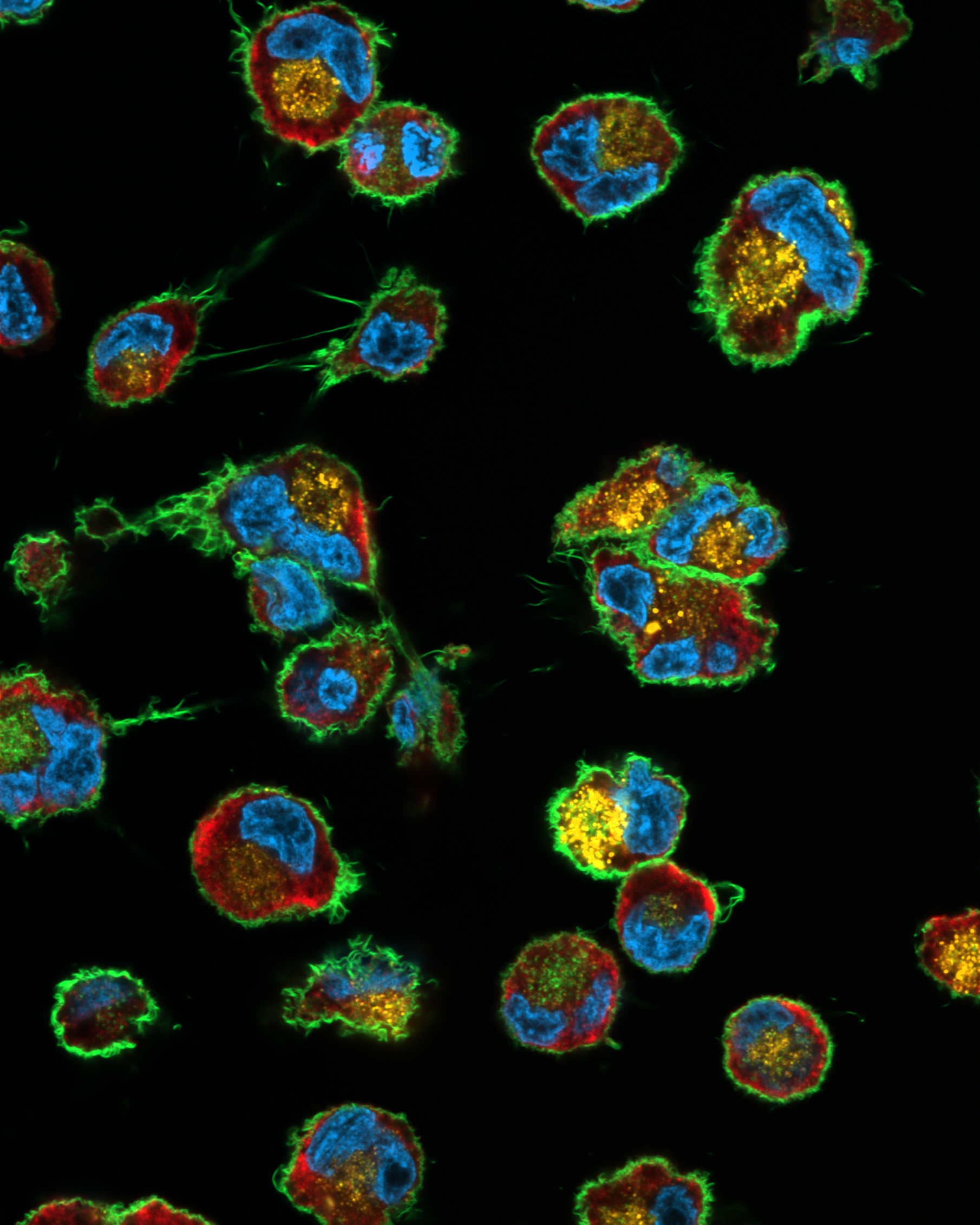
Due to their scalability and reproducibility, in vitro models of the intestine, alveolarepithelium, and endothelia (blood-brain barrier), represent a valuable test system during the optimization process of novel drug carriers. The flexibility provided through the use of cell lines further allows us to combine different cellular components of a tissue, depending on the desired complexity and intended read-out: Herein, we cultivate modular in vitro models that allow for the integration of different epithelial cells, fibroblasts, endothelial cells, as well as immune cells. Our established systems comprise a range of models, from standard CaCo-2 permeation testing to multi-layered in vitroskin models for investigating wound healing.
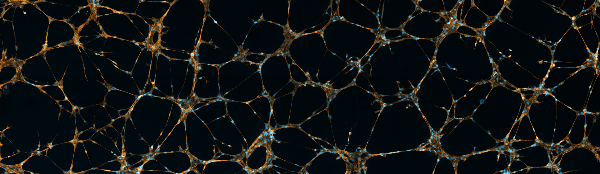
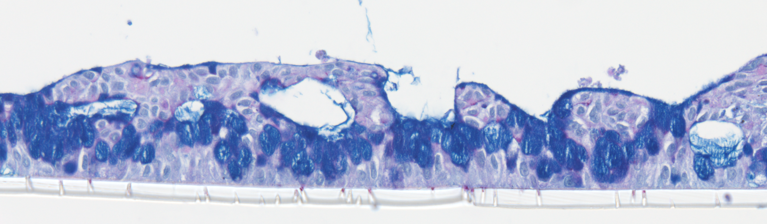
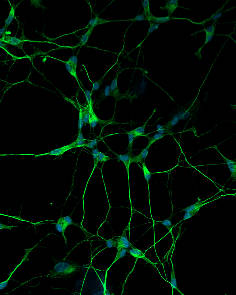
To answer scientific questions targeted at the molecular mechanisms and therapeutic strategies of inflammatory and infectious diseases, two-dimensional in vitro models often do not provide the necessary complexity of mimicking the diseased tissue microenvironment. In those cases, we rely on three-dimensional models, often cultivated on or within bio-inspired scaffolds and matrices to better replicate the respective physiological or pathological environment. Besides the use of established hydrogels for bioprinting and organoid cultivation, we develop functional materials to simulate the fibrillary structure of the native human extracellular matrix (ECM). Here, electrospun biomimetic scaffolds can be tuned in composition and elasticity to model, for example, the ECM of fibrotic or inflamed tissue in the lung and intestine. These scaffolds further provide optimal growth substrates for their integration into organ-on-chip (OoC) cultures.
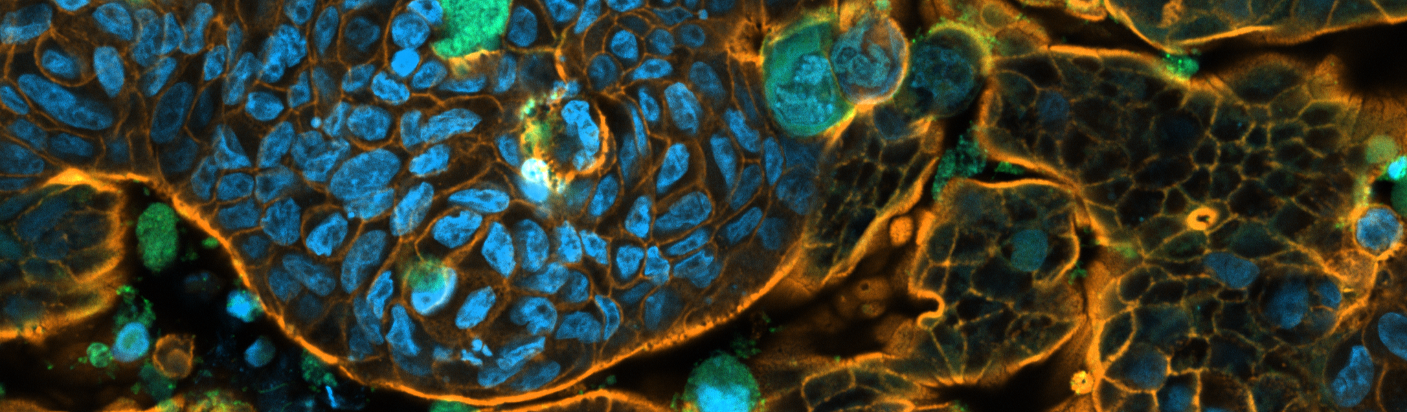
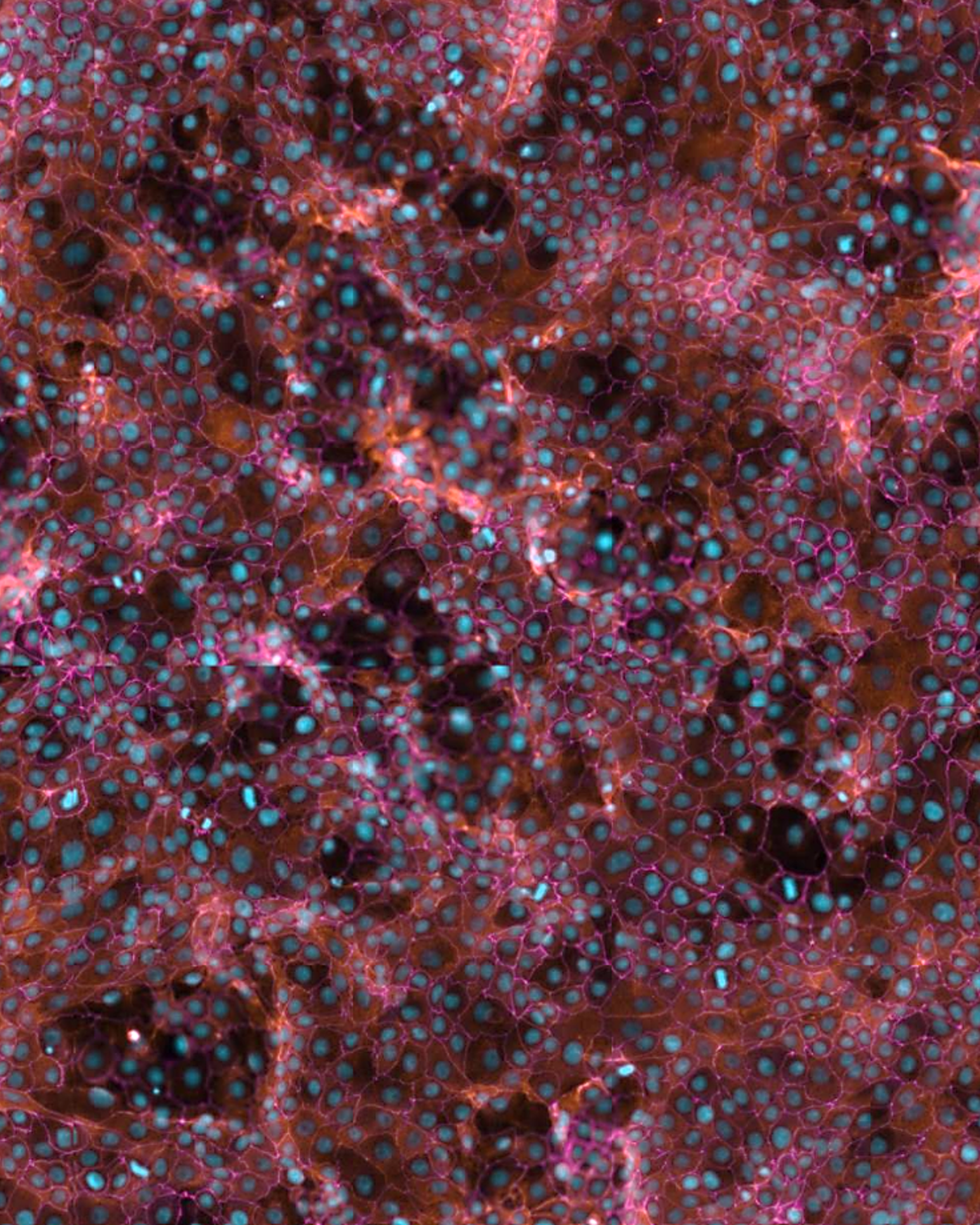
Although technological advancements now allow for the cultivation of human tissue cultures with complex architectures and multiple cell types, the use of living human tissue represents the gold standard in infection- and inflammation-centered research. Due to the complexity of tissue-resident immune components that are difficult to model in vitro, we employ ex vivohuman tissue to investigate molecular mechanisms associated with wound healing and host-pathogen interactions. Here, we specialize in the use of human skin to analyze the interaction of drug-delivery systems with healthy tissue and additionally evaluate the biological effects of formulations during wound healing.